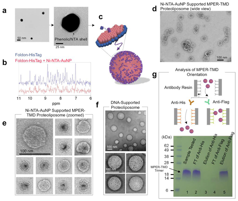
Figure 2. Nanoparticle-supported proteoliposome formation using bicelle-reconstituted MPER-TMD.
(a) The TEM images of gold-polyphenol nanoparticles (AuNPs) functionalized with NTA (NTA-AuNP). (b) 1H NMR spectra the Foldon protein with C-terminal His6-tag in the absence and presence of Ni-NTA-AuNPs. Blue: 450 μl of 30 μM Foldon-His6; Red: 450 μl of 30 μM Foldon-His6 mixed with 100 μl of Ni-NTA-AuNP (OD530 = 0.1) (the volume of the mixture adjusted to 450 μl before NMR measurement). (c) Schematic illustration of unidirectional coating of bicelle-reconstituted MPER-TMD onto Ni-NTA-AuNP. (d) Negative staining EM (nsEM) image (wide view) of Ni-NTA-AuNP supported MPER-TMD proteoliposome (see text). (e) nsEM images of the Ni-NTA-AuNP-supported MPER-TMD proteoliposomes at two magnifications. (f) nsEM images of DNA buckyball-supported MPER-TMD proteoliposomes at two magnifications. (g) Analysis of FLAG-MPER-TMD-His6 orientation in Ni-NTA-AuNP-supported liposomes by antibody resin pull-down and SDS-PAGE. Lane 1: Ni-NTA-AuNP-supported MPER-TMD liposome; Lane 2: flow-through from anti-His6 resin after 30 minutes incubation; Lane 3: elution from anti-His6 resin; Lane 4: flow-through from anti-FLAG resin after 30 minutes incubation; Lane 5: elution from anti-FLAG resin.

