
Figure 3.
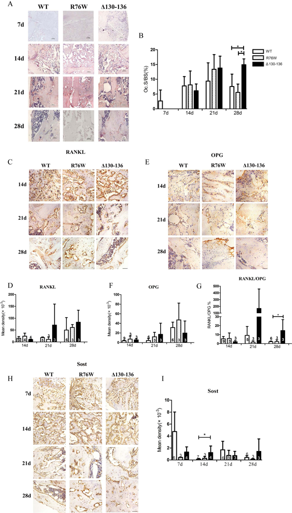
Blocking gap junctions delayed the bone repair process. (A) Representative images of TRAP staining in callus of WT, R76W and Δ130–136 mice at 7, 14, 21 and 28 days after fracture. (B) Quantification of TRAP+ osteoclasts (Oc.S/BS) in callus. Representative images of RANKL (C), OPG (E) and Sost (H) immunostaining in callus of WT, R76W and Δ130–136 mice after fracture. Quantification of RANKL (D), OPG (F), Sost (I) expression and ratio of RANKL/OPG (G). Data shown are mean ± SD. *p < 0.05. n=3–9. RANKL = Receptor Activator for Nuclear Factor-κB Ligand; OPG = osteoprotegerin; Sost = sclerostin. Scale bars = 50μm.