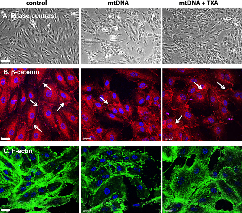
Figure 2.

TXA protects endothelial monolayer from damage induced by exogenous mtDNA. HUVEC growing on fibronectin coated glass coverslips were incubated for 1 h in serum-free medium with 0.3 μg/ml mouse mtDNA, in presence or absence of 100 μg/ml TXA. Cells were formalin fixed, fluorescently stained for β-catenin, actin cytoskeleton and nuclear DNA as described in Material and Methods and studied using a phase contrast microscope (A) or a confocal microscope (B and C). Bar in A – 70 μm. Bars in B and C – 23 μm.
