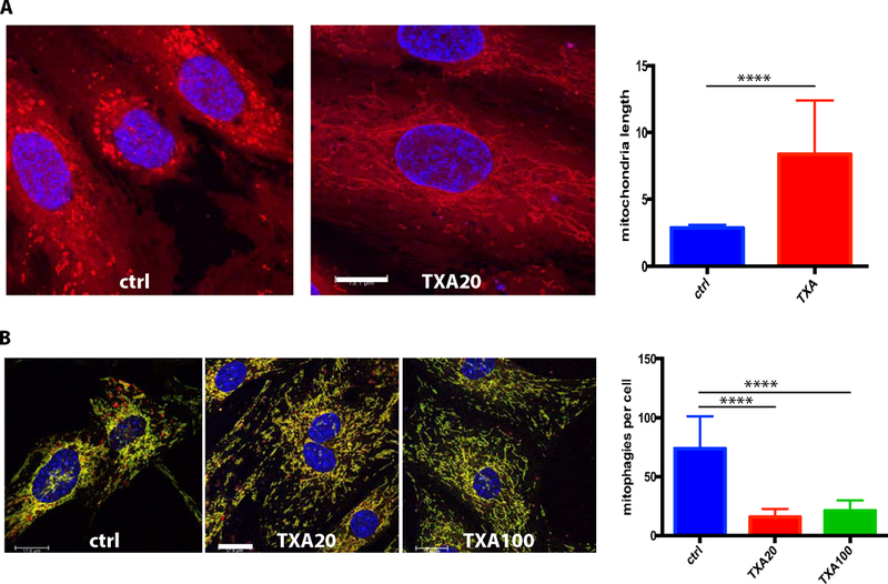
Figure 4. Effects of TXA on mitochondria.

A) HUVECs were treated for 24 h with 0 or 20 μg/ml TXA, stained with Deep Red Mitotracker, formalin fixed and studied using a confocal microscope. The mitochondria lengths were measured using the Image J program, and the mean mitochondria length and SD were calculated. TXA treated cells display longer mitochondria. Bar – 13.1 μm. B) Lung endothelial cells obtained from mito-QC mice were incubated for 24h in serum free medim with 0, 20 or 100 μg/ml TXA to assess mitophagy using confocal microscopy. The mean number of mitophagies (red only structures) per cell and SD were calculated. TXA decreases the presence of mitochondia that have underwent mitophagy (red only fluorescence). Bar – 18 μm.
