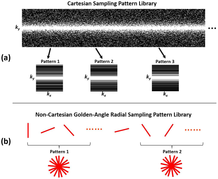
Figure 2.
Schematic demonstration of the undersampling patterns used in the study. (a) Examples of the 1D variable-density Cartesian random undersampling patterns used for knee imaging. (b) Example of the gold-angle radial undersampling masks used for liver imaging. The undersampling mask was varying for each iteration during the network training to augment the training data for SANTIS framework.

