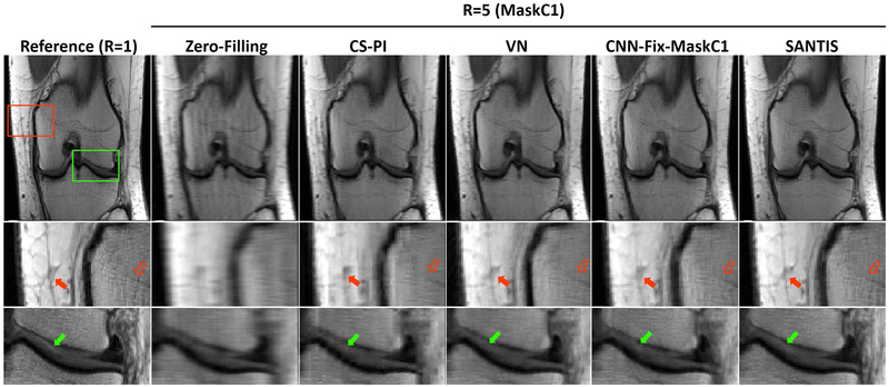
Figure 4.
Representative examples of knee images obtained using the different reconstruction methods at R=5. SANTIS showed the highest image quality with a better representation of the layered structure of the femoral and tibial cartilage (green arrows) with favorable preservation of tissue sharpness and texture (red arrows) comparable to the reference.

