. 2019 Jul 19;60(8):774–781. doi: 10.3349/ymj.2019.60.8.774

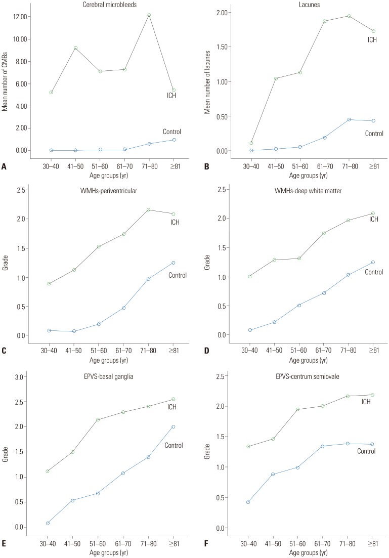
Fig. 2. The distribution of small vessel disease imaging markers by age group. Cerebral microbleeds (CMB) (A), lacunae (B), white matter hyperintensity (WMH) (C and D), and enlarged perivascular space (EPVS) (E and F) in the intracerebral hemorrhage (ICH) group and the control group. In all age groups, CMB, lacunae, WMH, and EPVS were more severe in the ICH group than in the control group. CMBs were prominent starting in the 30s and remained consistently high with aging in the ICH group. WMH in the periventricular area and deep white matter, EPVS in the basal ganglia and centrum semiovale, and lacunae all increased gradually with aging in both groups. These MRI markers had a consistently worse grade in the ICH group, even in young patients.

Fig. 2