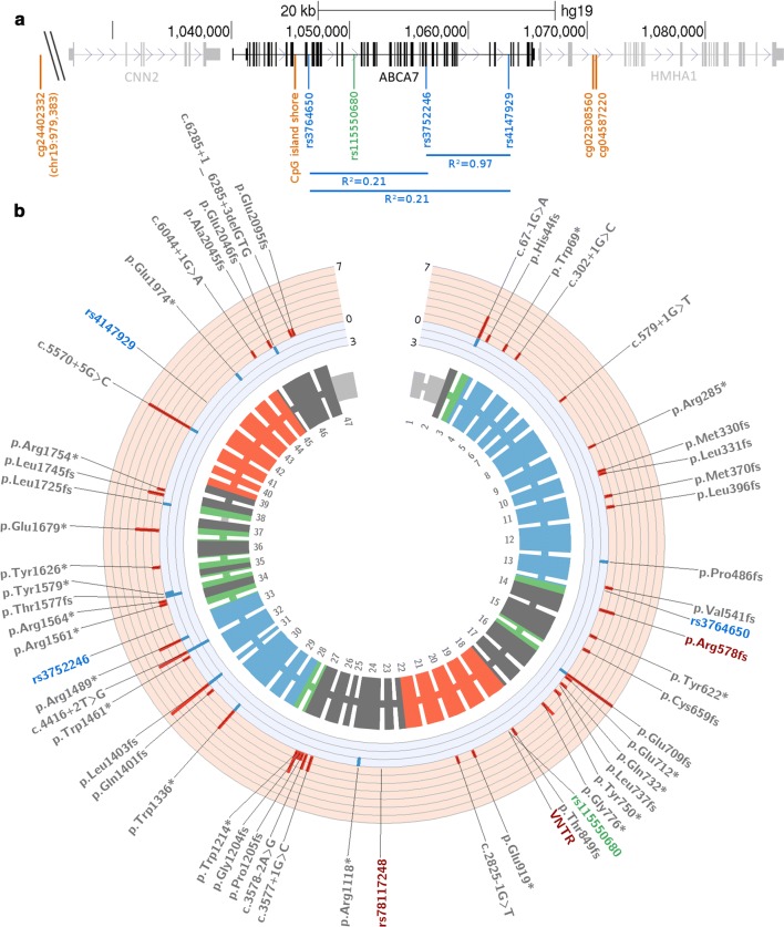
Fig. 1.
Genomic ABCA7 layout. a Part of the ABCA7 locus on chromosome 19 is shown (chr19:1026,000-1087,000 [hg19]), containing the ABCA7 gene, flanked by the CNN2 and HMHA1 genes. Significantly, AD-associated CpG methylation markers (orange), and GWAS sentinel SNPs from African American (green) and Caucasian (blue) study populations are shown, with R2 LD values for the latter. b Detailed ABCA7 plot, generated with circos [18]. From the outside to the inside: GWAS sentinel SNPs observed in African American (green), or Caucasian cohorts (blue), common functional variants (red), and PTC variants reported in AD case–control studies (gray) are shown. The subsequent track depicts the number of studies per PTC variant which report enrichment in AD (red, outward facing), or controls (blue, inward facing). The inner track corresponds to the 47 exons of ABCA7 with protein annotation: UTR (light gray), transmembrane domains (green), extracellular domains (blue), NBD domains (red), and unknown protein domains (dark gray)

