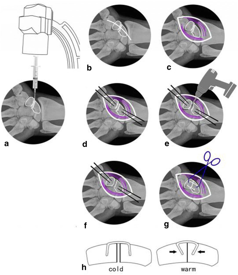
Fig. 3.
a–c Determine the location with a 5-ml syringe needle puncture under C-arm fluoroscopy and expose the scaphoid nonunion. d Proper locations for implantation were determined at proximal and distal poles of the bone and then, two φ1.2-mm K-wires were drilled in for poking. e Fibrous scar tissues and sclerotic calluses were removed through curetting using a scoop or drilling using an electric drill until bleeding spots were seen from the fractured margins. f The cancellous bone block graft with attached cortex was trimmed to match the size and shape of the deficits in the scaphoid; cancellous bone was used to adequately fill the treated proximal and distal fracture fragments followed by fracture reduction. Then, the graft of cancellous bone with cortex was embedded in the deficit site. g, h Choose the appropriate ASC and insert it, and then warm (35–40 °C) salt water was sprayed to gradually tighten the extended arms until their original shape was almost achieved

