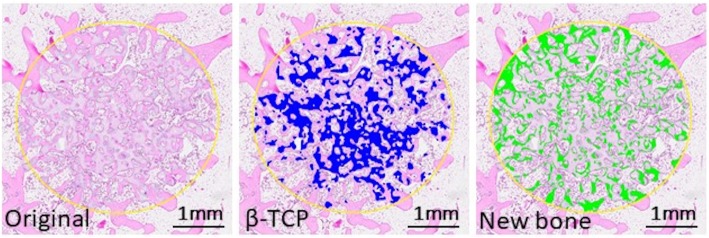
Fig. 2.
An image analysis of a bone defect. The surface area of newly formed bone and the remaining β-TCP was measured using an image analyzer, (WinROOF, Mitani Co., Tokyo, Japan). The colored area was used for evaluation of the residual amount of β-TCP (blue) and newly formed bone (green) in the defect. The scale bar shows 1 mm

