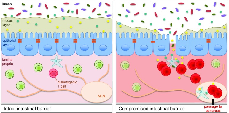
Fig. 1.
A compromised intestinal barrier triggers activation of diabetogenic T cells in the GALT. An intact intestinal barrier (Left) is composed of a thick mucus layer and an epithelial layer with tight junctions (orange). The rare diabetogenic T cells in the underlying lamina propria remain quiescent. In contrast, a compromised intestinal barrier (Right) characterized by a thin and discontinuous mucus layer and a breached epithelial barrier allows invasion of microbiota into the lamina propria. This results in activation and expansion of diabetogenic T cells in the GALT, which then travel via mesenteric (MLN) and pancreatic lymph nodes to the pancreas and induce diabetes.

