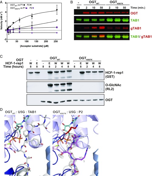
Fig. 2.
The N567K mutation abrogates OGT activity due to disrupted acceptor binding site. (A) Michaelis–Menten kinetics of OGT glycosyltransferase activity against P1 and P2 peptides. (B) Immunoblots showing OGT glycosyltransferase activity against TAB1. (C) Immunoblots showing OGT glycosyltransferase and proteolytic activities against HCF1-rep1. HCF1-rep1: GST-tagged host cell factor-1 fragment containing the first PRO repeat. W: wild type. E: E1019Q. (D) Crystal structures of OGT ternary complexes of OGTWT and OGTN567K active site in complex with UDP-5S-GlcNAc (U5G, turquoise, orange atoms) and acceptor peptide. OGTWT is shown in complex with RB2 (Retinoblastoma-like protein 2 peptide, light gray carbon atoms) [PDB ID code: 5C1D (57)]; OGTN567K is shown with P2 peptide (pink carbon atoms) (PDB ID code 6IBO). K567 is shown in red. An unbiased Fo–Fc difference map before inclusion of the peptide is shown as a mesh.

