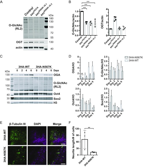
Fig. 3.
The N567K OGT mutation leads to altered O-GlcNAc homeostasis in D. melanogaster and neurite outgrowth defect in mESCs. (A) Immunoblot on 3- to 5-d-old adult Drosophila head lysates. (B) Quantification of O-GlcNAc modification on proteins and OGT protein level, normalized to actin signal. Protein O-GlcNAc modification is decreased in sxcN595K 19.1 (***P < 0.0001) and sxcN595K 10.3 (***P < 0.0001) lines to a comparable level found in the hypomorphic sxcH537A flies (***P < 0.0001). n = 4, mean ± SD. ANOVA with Tukey test. (C) Immunoblots on full cell lysate of 3HA-OGTWT and 3HA-OGTN567K mESCs showing OGA, Oct4, Sox2, Histone 3 (H3), and protein O-GlcNAcylation (RL2) levels. mESCs were differentiated for 0, 2, 4, and 6 d in N2B27 medium. (D) Quantification of immunoblots of OGA, Oct4, Sox2, and global protein O-GlcNAcylation in differentiated 3HA-OGTWT and 3HA-OGTN567K mESCs normalized to H3 signal. n = 4, mean ± SEM; **P = 0.012. Multiple t test using the Holm–Sidak method. (E) Immunofluorescence microscopy of 3HA-OGTWT and 3HA-OGTN567K mESCs after 8 d of neuronal differentiation in N2B27 medium. Cells were stained for tubulin III (green) and DAPI (magenta). (Scale bar, 50 μm.) (F) Quantification of neurite outgrowth. Total neurite length was measured per microscopic image by tracking β-tubulin III signal and then normalized to the number of nuclei (DAPI). (3HA-OGTWT, 108 to 800 nuclei per image; 3HA-OGTN567K, 260 to 740 nuclei per image) **P = 0.0079, Student t test, mean ± SD, n = 3.

