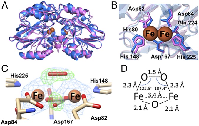
Fig. 5.
Crystal structures of Td and Tm ODP. (A) Overlay of the 2.07-Å resolution structure of iron-peroxo Td ODP (6R9N, blue) and the 2.0-Å resolution crystal structure of Zn-reconstituted Tm ODP (6QWO, magenta) (B) Di-iron centers of Td and Tm ODP conserve all metal-binding residues. (C) Di-iron site of Td ODP chain A contains a cis μ-1,2 iron-peroxo species and an oxo-bridge; 2Fo-Fc electron density map shown in blue at 2σ, and Fo-Fc omit map shown in green at 5.5σ. (D) Distances and angles of the iron-peroxo adduct are consistent with previously characterized cis-μ,1,2 iron-peroxo protein species and biomimetic compounds.

