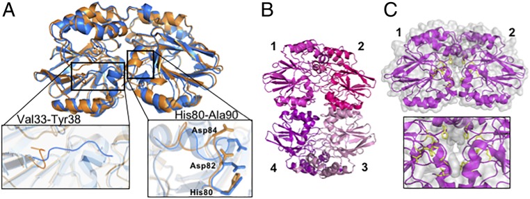
Fig. 6.
Crystal structures of apo Td and Tm ODP. (A) Apo Td ODP (6QNM, orange) and iron-peroxo Td ODP (6R9N, blue) have similar conformations, with the exception of 2 small loops composed of residues Val33-Tyr38 and His80-Ala90. Movement of these loops in the apo form exposes the active site to solvent. (B) Apo Tm ODP crystallizes as a tetramer (6QRQ), consistent with molecular weight measurements by MALS. Subunits 1 and 2 or 3 and 4 compose the dimer found in the metal-bound structure. (C) Solvent exposure of the active site residues (yellow) increases in the apo structure, and Trp102 from the adjacent subunit moves into the metal-free center (SI Appendix, Fig. S7E).

