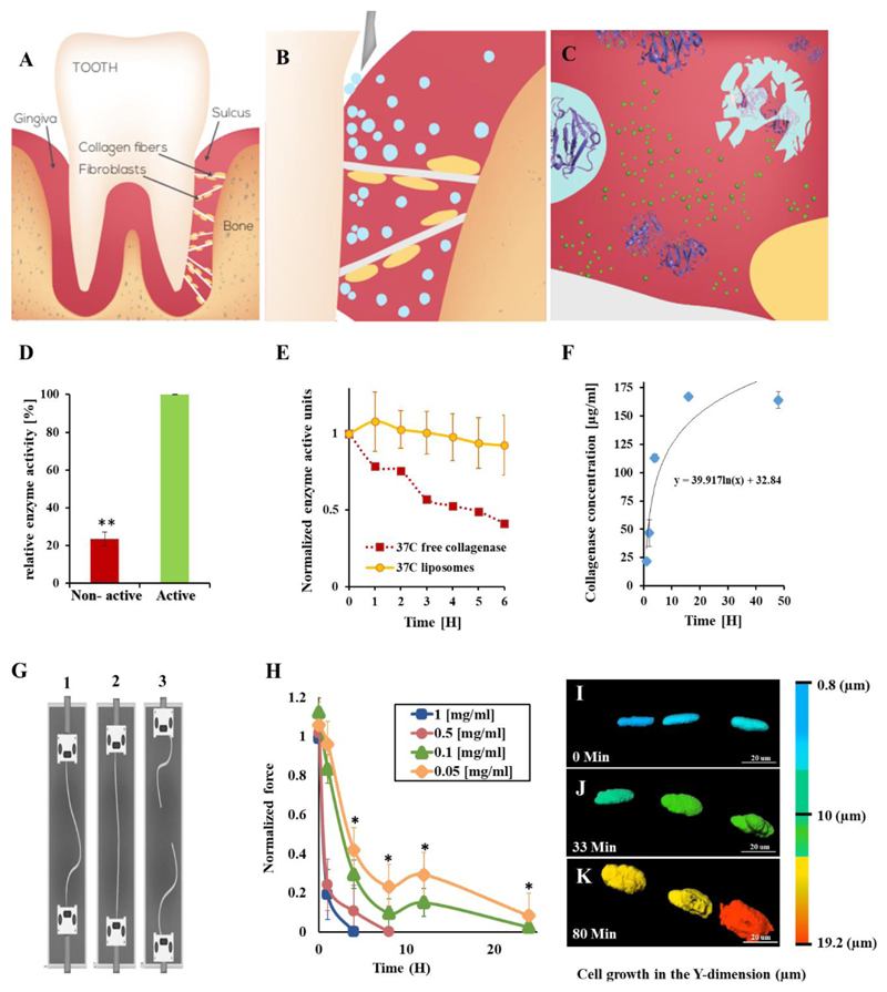
Figure 1. Nanosurgery – enzymes loaded into nanoparticles are released at the surgical site, where they are activated, to perform a localized surgical task.
Proteolytic enzymes, housed within nanoparticles replace the traditional blade, thereby, cutting only the target tissue, reducing pain and enhancing recovery. We utilized this approach to replace a surgical procedure in the oral tissue. Teeth are confined to their natural orientation by soft and hard tissue (A). Specifically, collagen type-1 fibers anchor the teeth to the underlying bone. Nanoparticles (blue spheres) loaded with collagenase, a proteolytic enzyme with specificity towards collagen, are inserted into the sulcus (B). The particles house the enzyme in a deactivated form. Once released, the Ca+2 naturally present in the tissue activates the enzyme and collagen degradation begins. The nanoparticles maintain the enzyme's therapeutic release profile and confine the biodistribution to the treatment site. (C) This facilitates enhanced orthodontic tooth movement. The collagen fibers recover rapidly after the treatment.
Controlled release and activation. Collagenase released from the nanoparticles over 48 hours at physiological conditions (F). Once released, the enzyme is activated by its cofactor calcium, naturally present in the tissue (D). The activity of the collagenase declines thereafter, over a period of 6 hours (E). As long as the enzyme is housed inside the particle it remains protected and inactive (D, and Supplementary Fig. S7A,B).
Collagenase relaxes collagen fibers. Collagen fibers were exposed to collagenase at various concentrations for different periods of time. The fibers were stressed using a force machine under oral physiological conditions (G, Supplementary Fig. S2, S7C). As the collagenase concentration increased, the fibers weakened (H). The higher collagenase concentrations (0.5 and 1 mg/mL) degraded the collagen fibers within less than 10 hours. A therapeutic window of 0.05 - 0.1 mg/ml at which the collagen fibers were relaxed but not fully degraded by collagenase was determined.
Fibroblast morphology changes as a function of collagenase activity. A collagen fiber (beneath the optical field) with adherent fibroblasts in focus was imaged during the process of collagenase treatment. As the collagen fiber relaxes, the adherent fibroblasts change their morphology from an elongated structure to a round structure (I→J→K, collagenase was added at t=0). A full time-lapse movie of the process is presented at: https://youtu.be/4krwecUS7Yc. It is important to note that the collagenase relaxed the collagen fibers but did not detach the fibroblasts nor impact their viability.
Error bars indicate the standard deviation of at least five independent measurements. ** indicates two tail P-Value <0.01.

