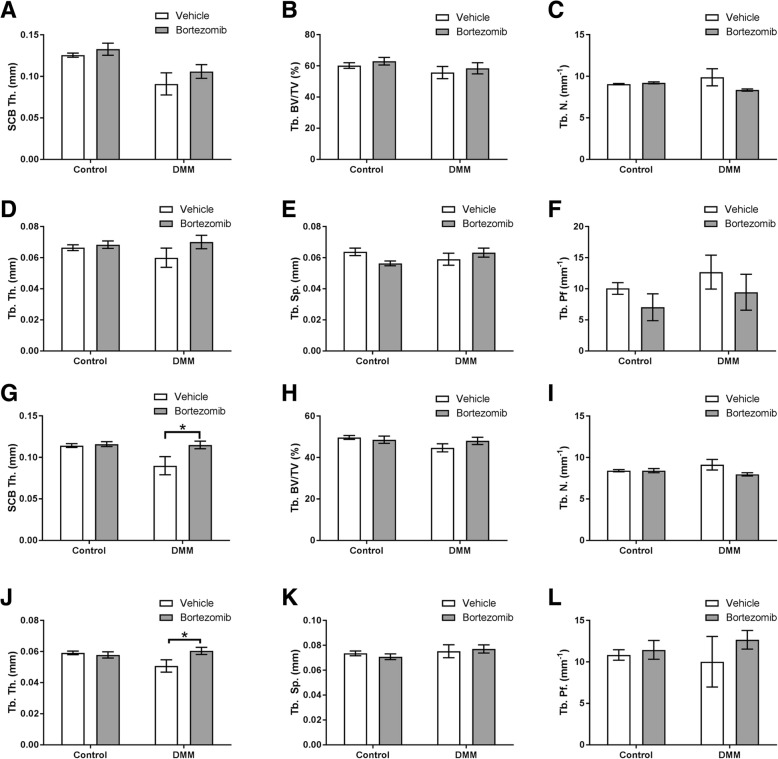
Fig. 3.
MicroCT analysis of the epiphyseal region of the medial tibia in DMM-operated and non-operated controls (a) subchondral bone thickness (SCB Th.) (b) trabecular bone volume/tissue volume (Tb. BV/TV) (c) trabecular number (Tb. N.) (d) trabecular thickness (Tb. Th.) e trabecular separation (Tb. Sp.) f trabecular pattern factor (Tb. Pf.). MicroCT analysis of the epiphyseal region of the lateral tibia in DMM-operated and non-operated controls (g) subchondral bone thickness (SCB Th.) (h) trabecular bone volume/tissue volume (Tb. BV/TV) (I) trabecular number (Tb. N.) (J) trabecular thickness (Tb. Th.) (k) trabecular separation (Tb. Sp.) (l) trabecular pattern factor (Tb. Pf.). Data are presented as mean ± S.E.M (n = 8/group). P < 0.05*

