Fig. 4.
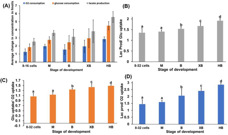
(A) Oxygen and glucose consumptions and lactate production of bovine embryos at various stages. (B) Flux ratio between lactate production and glucose uptake (Lac prod/Glu uptake). (C) Flux ratio between glucose and oxygen uptakes (Glu uptake/O2 uptake). (D) Flux ratio between lactate production and oxygen consumption (Lac prod/O2 uptake). In all figures: mean ± SD; dead oocytes or embryos (n= 12), 8 to 32 cells (n= 12), morula (M, n=7), blastocyst (B, n= 6), expanded blastocyst (XB, n= 17), and hatched blastocyst (n=8, HB). a, b, c, d within columns: Tukey HSD (pairwise comparison), values with different superscripts are significantly different (P≤ 0.001).

