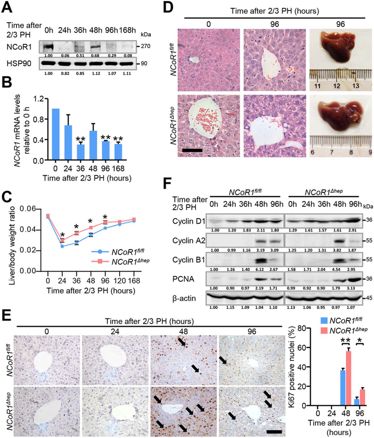
FIG. 1.
Liver regeneration after 2/3 partial hepatectomy is enhanced in NCoR1Δhep mice. (A) Protein and (B) mRNA expression change of NCoR1 in the process of liver regeneration were detected by western blot and qPCR, Data are presented as mean ± SEM (n=4). *P<0.05; **P<0.01. (C) Liver weight to body weight ratio analysis in NCoR1fl/fl and NCoR1Δhep mice over a time course from 0 to 168 hours after 2/3 PH. Data are presented as mean ± SEM (n=5). *P<0.05; **P<0.01, NCoR1Δhep mice compared to NCoR1fl/fl mice at the indicated times. (D) Morphological changes of livers of NCoR1fl/fl mice and NCoR1Δhep mice at the 96 hours after 2/3 PH as determined by H&E staining. scale bar: 50 μm. (E) Immunohistochemical analysis of Ki67 in paraffin tissues from livers of NCoR1fl/fl and NCoR1Δhep mice at the indicated times after 2/3 PH (Left). Quantification of the percentage of Ki67 labeled nuclei (Right). Data are presented as mean ± SEM (n=4). The different degrees of significance were indicated as follows in the graphs: *P<0.05, **P<0.01, NCoR1Δhep mice compared to NCoR1fl/fl at indicated time. (F) Western blot analysis of CyclinD1, CyclinA2, CyclinB1, PCNA using RIPA extracts of NCoR1fl/fl and NCoR1Δhep livers obtained at the indicated times after 2/3 PH. (Two Way ANOVA plus Student’s t test for B, C; Student’s t test for E).

