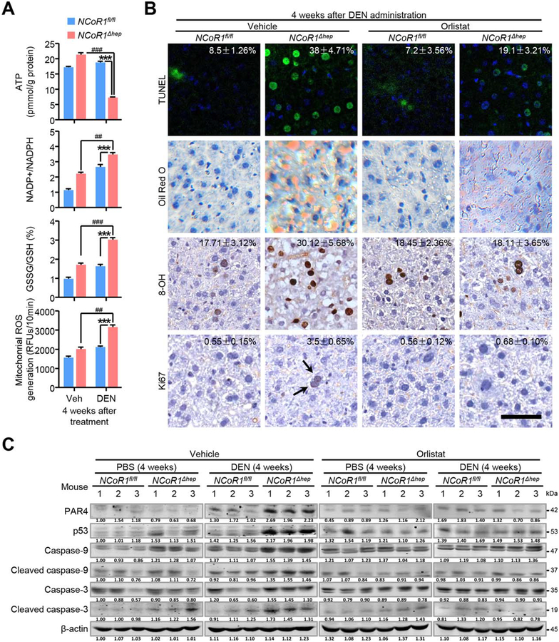
FIG. 6.
Increased oxidative stress and apoptosis in early stage of DEN injury in NCoR1Δhep mice. (A) Biochemical detection of intracellular ATP levels, NADP+/NADPH ratios, GSSG/GSH ratios and mitochondrial ROS in NCoR1fl/fl and NCoR1Δhep mice treated with PBS or DEN at four weeks after DEN treatment. Data are presented as mean ± SEM (n=4). ***P<0.001, NCoR1Δhep mice treated with DEN compared to NCoR1fl/fl mice treated with DEN. ##P<0.01; ###P<0.001, NCoR1Δhep mice treated with DEN compared to NCoR1Δhep mice treated with control vehicle. (B) Four weeks after DEN administration, NCoR1fl/fl and NCoR1Δhep mice were treated with vehicle or orlistat for 5 days before sample collection. Representative Tunel staining, Oil Red O staining and immunohistochemical analysis of 8-OH and Ki67 were performed on mice liver samples. Quantification of the percentage of Tunel, 8-OH and Ki67 labeled nuclei were marked. Data are presented as meaňSEM of at least four mice per group. scale bar: 50 μm. (C) Four weeks after DEN administration, NCoR1fl/fl and NCoR1Δhep mice were treated with vehicle or orlistat for 5 days. Western blot analysis of expression of protein level of p53, PAR4, Caspase-3 and Caspase-9 (full length and their cleaved form) in mice liver samples were performed. (Student’s t test for A, B).

