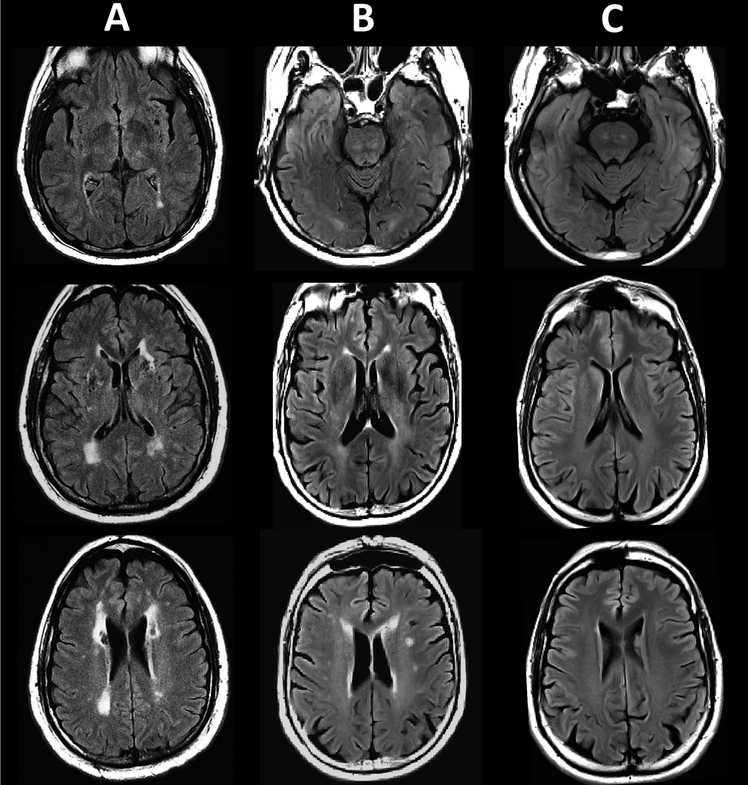
Figure 1.
Fluid-attenuated inversion recovery (FLAIR) images of white matter signal abnormalities in: A) a 61 year old male subject with CADASIL (image courtesy of Dr. Anand Viswanathan, J. Philip Kistler Stroke Research Center, Massachusetts General Hospital, Boston, USA); B) a 58 male subject with sporadic cerebral small vessel disease (SVD) and C) a 58 male subject with normal appearing white matter.

