Figure-3.

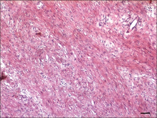
Hematoxylin and eosin stained section of the fibroma showing streams and bundles of well-differentiated spindle connective tissue cells that are haphazardly arranged and running in different directions with abundant collagen (Bar=50 µm).

Hematoxylin and eosin stained section of the fibroma showing streams and bundles of well-differentiated spindle connective tissue cells that are haphazardly arranged and running in different directions with abundant collagen (Bar=50 µm).