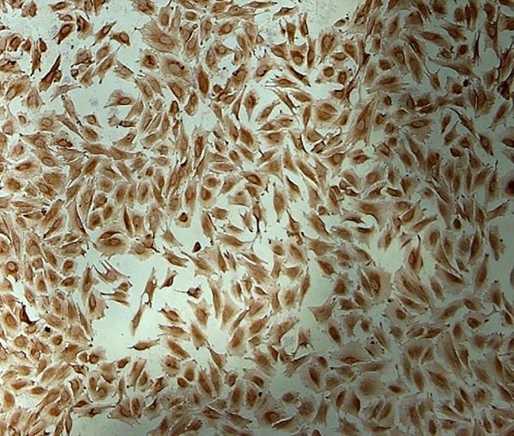
Figure1.
Immunocytochemistry analysis shows endometrial stromal fibroblasts. The cells were isolated from ectopic and eutopic endometrial tissues followed by staining with primary antibody against vimentin (10x magnification). Cells were observed under a phase-contrast microscope, and the representative field was photographed.

