Fig. 1.
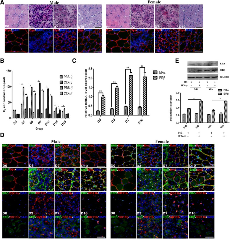
The alternation of serum estrogen level and estrogen receptor expression in CTX-damaged muscle. a Histological features of the inflamed TA muscle of male and female mice. The top images: standard H&E staining, and the bottom images: Dystrophin immunofluorescence staining (Red). b Elisa assay showing serum E2 levels. c qRT-PCR analysis presented the mRNA levels of estrogen receptor (ERs) gene in damaged TA muscle. d Representative immunofluorescence double-staining results of ERα/ERβ and Dystrophin, or ERα/ERβ and CD11b in damaged TA muscle. Aggregation of ERβ in myofibers was indicated by asterisk (*). e Western blots showing protein levels of ERα and ERβ in horse serum-differentiated C2C12 cells with or without IFN-γ treatment. The relative band intensities from western blots experiments were normalized to the level of GAPDH and analyzed with ImageJ software. All data are presented as mean ± SD (n = 3). One-way ANOVA was used for multiple comparisons. (*p < 0.05 and **p < 0.01). HS horse serum. Bar = 100 μm

