Fig. 3.

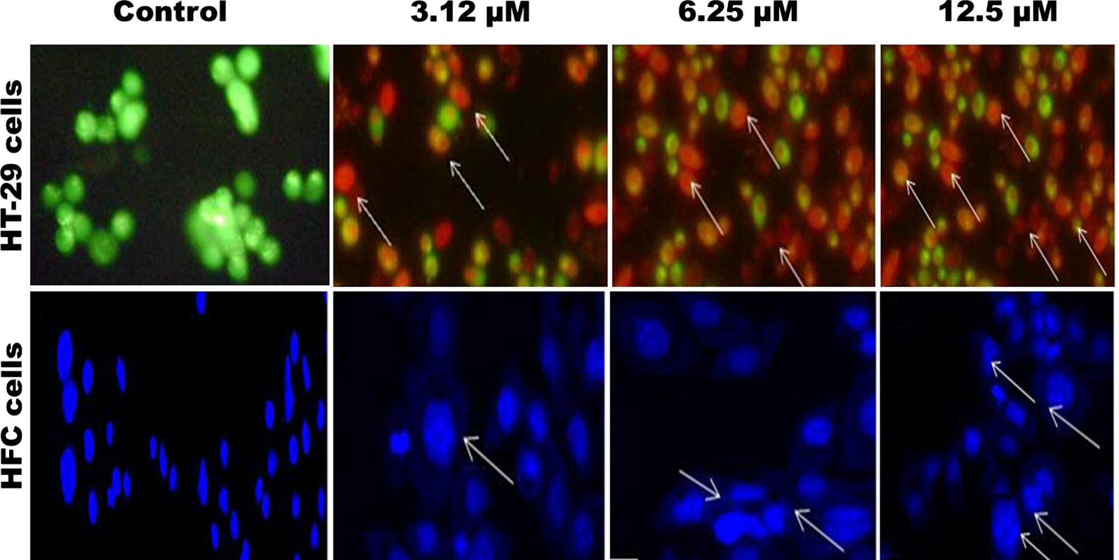
Nuclei morphological changes seen in HT-29 cells stained with AO/EtBr fluorescence microscopy. HT-29 cells were seeded in 6-well plates (4 × 10³ cells/mL) and treated with PAC compound with GI50 concentration respectively for 24 h at 37 °C

Nuclei morphological changes seen in HT-29 cells stained with AO/EtBr fluorescence microscopy. HT-29 cells were seeded in 6-well plates (4 × 10³ cells/mL) and treated with PAC compound with GI50 concentration respectively for 24 h at 37 °C