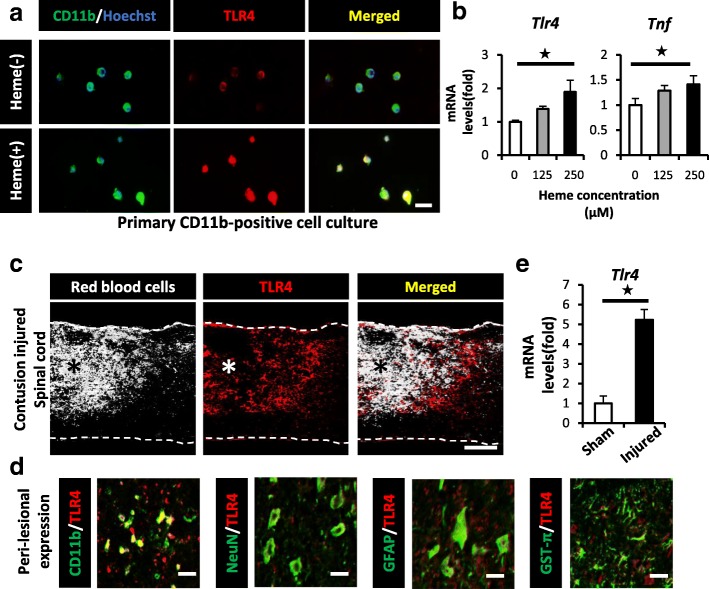
Fig. 4.
Heme exacerbates the TNF expression in CD11b-positive cells via the TLR4 pathway after SCI. a IHC showed heme-induced morphological changes and the expression of TLR4 (red) in primary CD11b-positive cells (green) at 30 min after heme stimulation (125 μM). b A qRT-PCR showed that the mRNA expression levels of TLR4 and TNF were both upregulated in a heme concentration-dependent manner (n = 4 in each group, triplicate). c IHC showed that intralesional bleeding (white) merged with TLR4-positive cells (red) in the peri-lesional area at 1 dpi. d IHC showed that CD11b-positive cells predominantly expressed TLR4 (red) around the lesion after SCI. CD11b, microglia; NeuN, neuron marker; GFAP, astrocyte marker; GST-π, oligodendrocyte marker (green). e The TLR4 mRNA expression in injured spinal cord at 1 dpi was significantly upregulated in comparison to sham spinal cord (n = 4 in each group). Scale bars, 20 μm (a, d), 500 μm (c). *p < 0.05, Wilcoxon rank-sum test (e) and Dunnett’s test in comparison to 0 μM heme-stimulation (b). Error bars indicate the SEM

