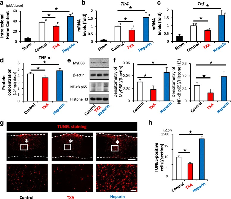
Fig. 5.
TXA alleviates the intralesional heme content, TLR4/TNF axis, and the number of apoptotic cells after SCI. a The intralesional heme concentration in injured spinal cords was increased in comparison to sham spinal cords at 1 dpi (#) (n = 6 in each group). The administration of TXA significantly reduced the heme content (black star). b, c A qRT-PCR showed that the administration of TXA significantly downregulated both the TLR4 and TNF mRNA expression in injured spinal cord at 1 dpi (n = 4 in each group). d Protein concentration of TNF-α of TXA-treated mice was significantly reduced compared to saline-treated mice at 3 dpi (n = 9 in each group). e, f Densitometric scanning of the immunoblots of MyD88, and NF-κB p65 (n = 9 per group). *p < 0.05, Dunnett’s test. The error bars indicate the SEM. g TUNEL staining showed the presence of TUNEL-positive apoptotic cells (red) around the lesion epicenter at 1 dpi. Bottom figures show higher magnification views corresponding to the boxed areas in the top figures. h Quantification of e demonstrating that the administration of TXA significantly reduced the number of TUNEL-positive apoptotic cells (n = 6 in each group). Scale bars, 500 μm (e left), and 50 μm (e right). *p < 0.05, Dunnett’s test (a–d, f, h) in comparison to control mice (black star) and sham-treated (laminectomy only) mice (#). Error bars indicate the SEM

