
Fig. 5.
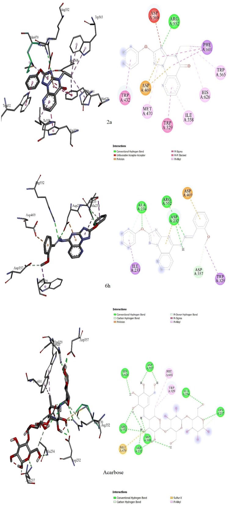
Molecular docking comparison of acarbose with compounds 2a and 6h. 3D binding conformation (right) and 2D binding conformation (left) showing the closest interactions between the active site residues of α-glucosidase and the most active (6h), least active (2a) synthesized derivatives and acarbose