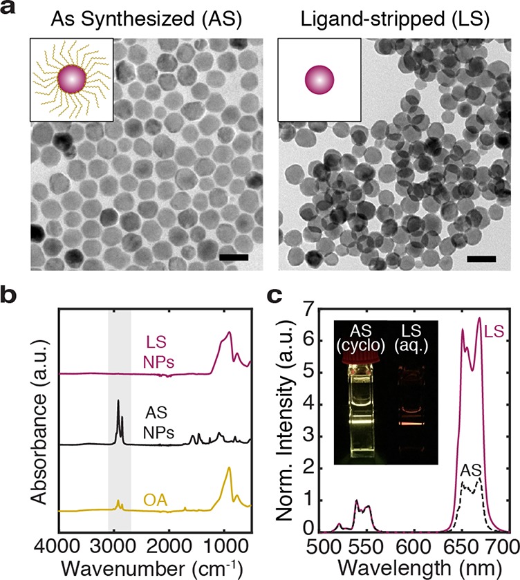
Figure 1.

Upconverting nanoparticles before and after ligand-stripping. (a) Core–shell (NaYF4:Yb,Er@NaLuF4) nanoparticles are as synthesized (AS) with an oleic acid (OA) coating and ligand-stripped (LS) for dispersion in aqueous media. Transmission electron micrographs (TEMs) show the quasispherical morphology and monodispersity of AS nanoparticles and LS nanoparticles. The scale bars are 50 nm. (b) Fourier-transform infrared spectroscopy (FTIR) measurements of OA (gold), AS nanoparticles (black), and LS nanoparticles (maroon) confirm that the highlighted vibrational modes around 2900 cm–1, associated with organic molecules like OA, are no longer present after the ligand-stripping procedure. Here, each spectrum is normalized to its maximum peak. For LS NPs and OA, the dominant peak around 910 cm–1 comes primarily from the glass slide (see Figure S1). (c) Upconversion spectra of AS nanoparticles in cyclohexane (black) and LS nanoparticles in an aqueous medium, S-Medium (maroon). Each spectrum is normalized to its green emission peak. The inset qualitatively shows the corresponding emission of colloidally suspended nanoparticles in a cuvette under 980 nm laser illumination. Enhanced contribution of the red emission band explains the perceived color difference between AS and LS UCNPs.
