Figure 1.

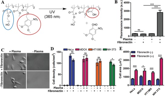
Characteristics of fibronectin‐coated PDMP. A) Photochemical reaction of PDMP. Blue circle: PEG side chain, red circle: organic soluble group (left side) to water soluble group (right side) conversion by the photochemical reaction. B) Effects of plasma treatment on the fibronectin adsorption on PDMP surfaces. The amount of fibronectin on the PDMP surfaces was measured by immunofluorescence microscopy. Fluorescence intensity is arbitrary unit (a.u.). C–E) Effects of plasma treatment and fibronectin coating of PDMP surfaces on cell adhesion. Cells seeded on each type of the surface were incubated for 3 h, washed to remove unbound cells, and DIC images were acquired. Representative DIC images of HeLa cells on different types of surfaces are shown in C (Scale bar: 50 µm). Using DIC images, D) cell density and E) cell area of four different cells (HT1080, MDCK, HeLa, and NIH 3T3) were measured on various types of surfaces. Data are shown as mean ± s.e.m. [two‐sided Student's t‐test] ns: not significant, *p < 0.05, *** p< 0.001.
