Figure 3.

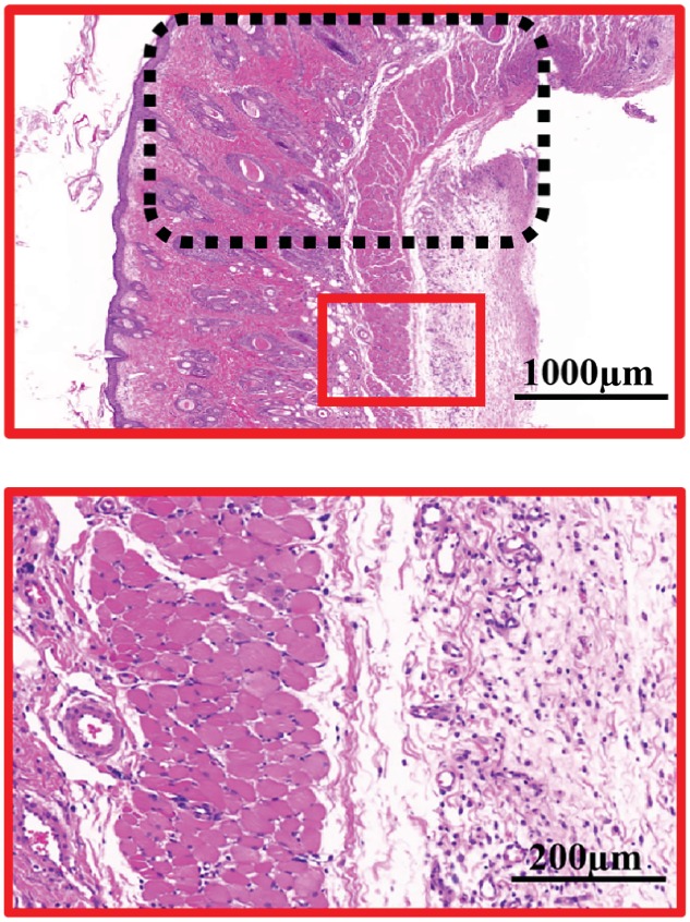
e, third row) H&E staining of the necrosis and survival junction area of the skin flaps in the 40 μM MF‐Lip treated sample. e, last row) Magnified view of the survival area in the 40 μM MF‐Lip treated sample. *p < 0.05.

e, third row) H&E staining of the necrosis and survival junction area of the skin flaps in the 40 μM MF‐Lip treated sample. e, last row) Magnified view of the survival area in the 40 μM MF‐Lip treated sample. *p < 0.05.