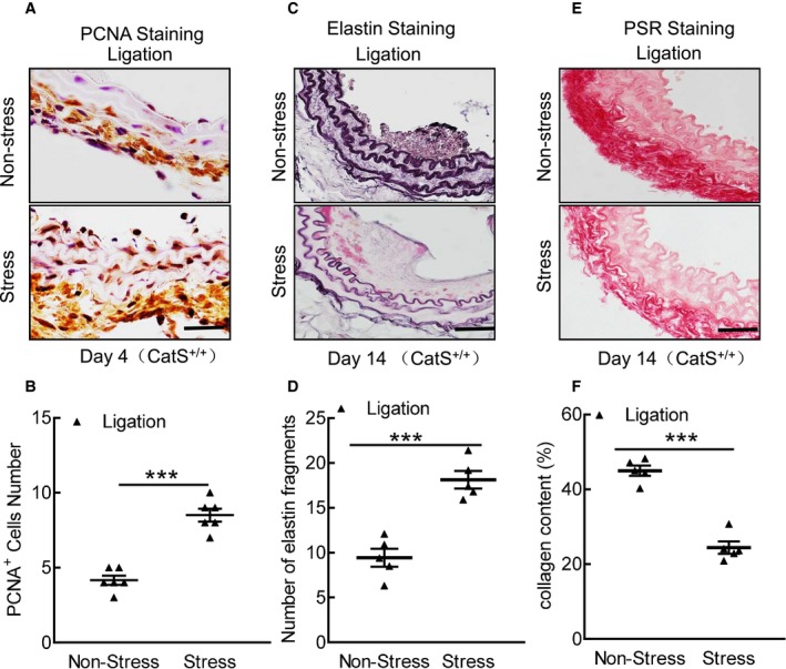
Figure 2.

Stress accelerates cell proliferation and media elastic laminal degradation and collagen in cathepsin S wild‐type (CatS+/+) mice. A and B, Representative proliferating cell nuclear antigen (PCNA) immunostaining images of media smooth muscle cell proliferation and combined quantitative data for PCNA + cells. C through F, Representative images and quantitative data for the number of broken elastin fragments (Elastica van Gieson) and collagen content (picrosirius red [PSR]). Bar=100 μm. The results are mean±SEM (n=5–6). ***P<0.001 vs CatS+/+ group by Student t test.
