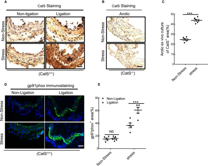
Figure 5.

Immunostaining showed the cell sources of cathepsin S (CatS) in injured arteries. A and B, Representative images of CatS staining that show the positive staining signaling was pronounced in the media and neointima (thin) of injured vessels from wild‐type CatS (CatS+/+) mice on day 4 after surgery, whereas no signaling was detected in the whole arterial walls of CatS‐deficient (CatS−/−) mice. C, CatS staining of cross‐sections taken on day 4 after ex vivo cultured aortae of CatS+/+ mice received stress. D and E, Representative images and quantitative data show the gp91phox+ staining area. Results are mean±SEM (n=5–7). NS indicates no significance. ***P<0.001 vs nonstressed group by 1‐way ANOVA, followed by Tukey post hoc tests.
