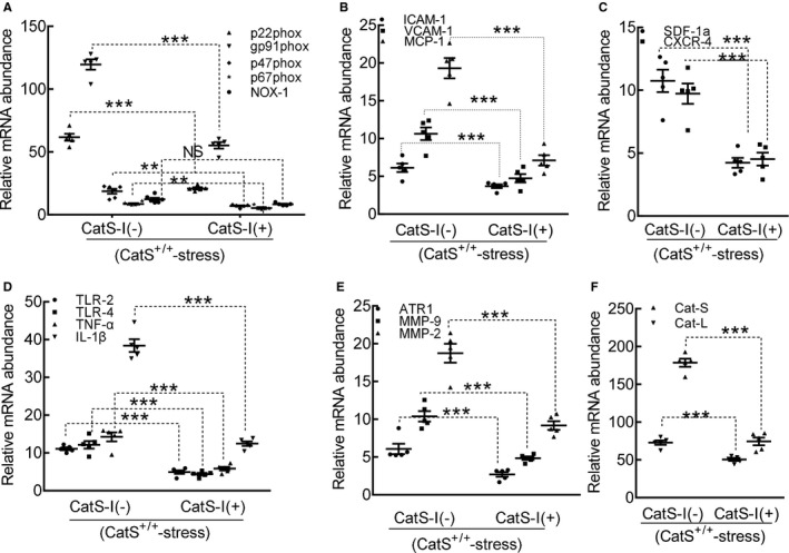
Figure 16.

Cathepsin S inhibition (CatS‐I) mitigated the expressions of targeted oxidative stress–, inflammation‐, and proteolysis‐related genes in the carotid arteries of the stressed CatS wild‐type (CatS+/+) mice at day 4 after surgery. A through F, Quantitative polymerase chain reaction data show the levels of p22phox, gp91phox, p47phox, p67phox, nicotinamide‐adenine dinucleotide phosphate, reduced form, oxidase 1 (NOX‐1), intercellular adhesion molecule‐1 (ICAM‐1), vascular cell adhesion molecule‐1 (VCAM‐1), monocyte chemoattractant protein‐1 (MCP‐1), stromal cell–derived factor‐1α (SDF‐1α), C‐X‐C chemokine receptor‐4 (CXCR‐4), toll‐like receptor (TLR)‐2, TLR‐4, tumor necrosis factor (TNF)‐α, interleukin‐1β (IL‐1β), angiotensin II receptor 1α (ATR1α), matrix metalloproteinase (MMP)‐9, MMP‐2, cathepsin (CatL), and CatS mRNAs of mice with CatS‐I (CatS‐I [−]) and mice with CatS‐I (CatS‐I [+]). Results are mean±SEM (n=5–7). NS indicates no significance. **P<0.01/***P<0.001 vs corresponding CatS‐I (−) mice by 1‐way ANOVA, followed by Tukey post hoc tests.
