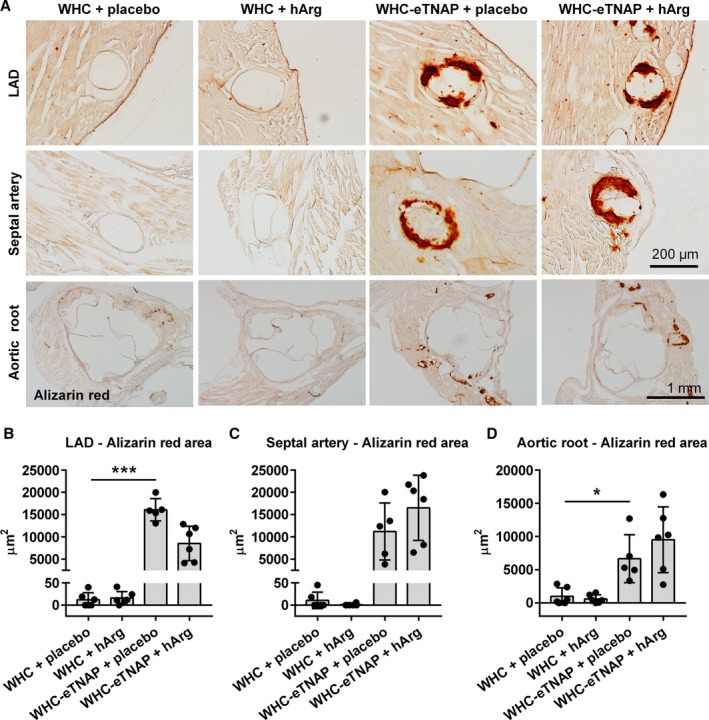
Figure 4.

Alizarin red staining for calcium. A, Left anterior descending artery (LAD, top panels), proximal segment of the septal artery (middle panels), and the aortic root (bottom panels). B through D, Morphometric quantification of alizarin red positive area in the LAD (B), septal artery (C), and the aortic root (D). *P<0.05, ***P<0.001. eTNAP indicates overexpression of tissue‐nonspecific alkaline phosphatase in the endothelium; hArg, homoarginine; LAD, left anterior descending artery; WHC, wicked high‐cholesterol allele.
