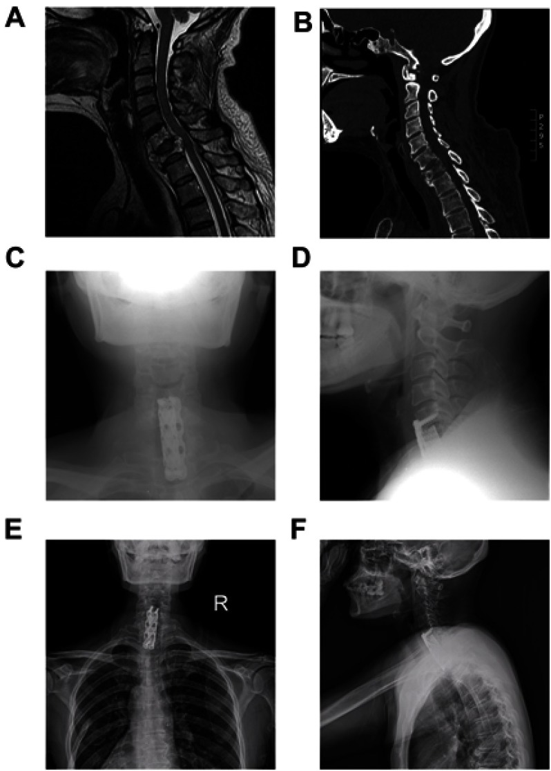
Figure 3.
Radiographic images of a representative 55-year-old female patient (Case #15).
Notes: Preoperative MRI (A) and CT (B) showing destruction of C6-7 vertebrae with instability and spinal cord compression. (C-D) Postoperative radiograph after total piecemeal spondylectomy and internal fixation using titanium mesh and plate osteosynthesis system. (E-F) X-ray image at 23 months after surgery showing no local relapse and stable instrumentation.
Abbreviations: MRI, magnetic resonance imaging; CT, computed tomography.

