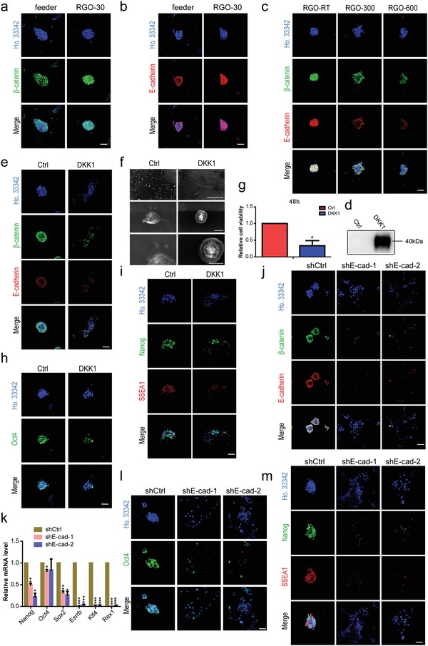
Figure 7.

RGO substrates activate β‐catenin/E‐cadherin expression in maintaining ESC pluripotency. The immunofluorescent staining of a) pluripotency‐maintaining protein β‐catenin and b) cell adhesion–associated protein E‐cadherin of 46C cultured on feeder and RGO‐30 substrates. Scale bar: 50 µm in panels (a) and (b). c) The protein expression levels of β‐catenin (green) and E‐cadherin (red) in 46C cultured on RGO‐RT, RGO‐300, and RGO‐600 substrates. Scale bar: 50 µm. d) Western blotting of RGO‐30 substrates after being modified by water and DKK1, respectively. Dickkopf related protein 1 (DKK1) was the inhibitor of the classic Wnt signaling pathway. Water‐modified substrate was used as a control (Ctrl). e) The protein expression levels of β‐catenin (green) and E‐cadherin (red) in 46C cultured on water or DKK1‐modified substrate. Scale bar: 50 µm. f) The SEM images with different magnifications of a single cell of 46C cultured on water or DKK1‐modified substrate for 4 h. Magnifications: 150 × (the upper line), 5000 × (the middle line), and 10 000 × (the lower line). Scale bars: 300 µm in the upper line, 5 µm in the middle line, and 4 µm in the lower line. g) The relative cell viability of 46C cultured on water or DKK1‐modified substrate for 48 h was detected by CCK‐8 kit. * p < 0.05 versus water‐modified substrate. The protein expression levels of h) Oct4, i) Nanog and SSEA1 in 46C cultured on water or DKK1‐modified substrate were detected by immunofluorescent staining. Scale bar: 50 µm in panels (h) and (i). The protein levels of j) β‐catenin and E‐cadherin of shCtrl, shE‐cad‐1, and shE‐cad‐2 on RGO‐30 substrate. Scale bar: 50 µm. k) The expression levels of Nanog, Oct4, Sox2, Esrrb, Klf4, and Rex1 of shCtrl, shE‐cad‐1, and shE‐cad‐2 cultured on RGO‐30 substrate were detected by qRT‐PCR. * p < 0.05, ** p < 0.01, *** p < 0.001 versus the shCtrl group. The representative fluorescence microscope images of l) Oct4, m) Nanog, and SSEA1 expressed by shCtrl, shE‐cad‐1, and shE‐cad‐2 on RGO‐30 substrate. Scale bar: 50 µm.
