Figure 11.

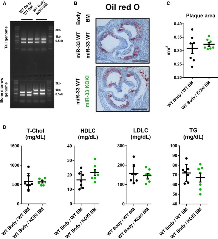
Hematopoietic microRNAs (miR‐33a and miR‐33b) have similar effects on atherosclerotic plaque formation. A, Representative genotyping polymerase chain reaction analysis of tail and blood cell genome. B, Representative microscopic images of the proximal aorta in apolipoprotein E–deficient (ApoE−/−)/miR‐33 wild‐type (WT) mice transplanted with bone marrow (BM) obtained from ApoE−/−/miR‐33 WT mice or ApoE−/−/miR‐33 knock out knock in (KOKI) mice. Bar=500 μm. C, Quantification of the atherosclerotic plaque area in cross‐sections at the proximal aorta level. D, Serum lipid profiles of ApoE−/−/miR‐33 WT mice transplanted with BM obtained from ApoE−/−/miR‐33 WT mice or ApoE−/−/miR‐33 KOKI mice. n=7 for each. Horizontal bars in the dot plots indicate mean±SEM. HDLC indicates high‐density lipoprotein‐cholesterol; LDLC, low‐density lipoprotein‐cholesterol; T‐Chol, total cholesterol; TG, Triglyceride.
