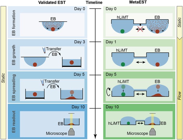
Figure 1.

Concept of metaEST. Side‐by side comparison of the metaEST concept and the validated standard EST. EBs (red) from mouse embryonic stem cells are formed at the liquid–air interface of a hanging drop under static conditions. During EB formation, the EB compartment of the metaEST is separated from the compartment hosting human liver microtissues (hLiMTs, green), both of which are on the same chip. At day 1 of the metaEST, hLiMTs and EBs are fluidically interconnected in the same network, while the validated EST is continued under static and monoculture conditions. In the EST, the EBs are then transferred to suspension culture on day 3 to replenish the medium. In the metaEST, this transfer is not necessary, as medium is exchanged directly on‐chip. At day 5, the EBs in the validated EST are manually transferred to an adhesive surface, where they adhere and spread out. In the metaEST this step is realized by flipping the microfluidic chip upside down to a standing‐drop configuration so that the substrate of the chip serves as a substrate for EB spreading. At day 10, the EBs are morphologically inspected under the microscope to detect beating areas in the spread tissues.
