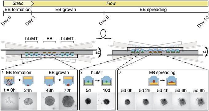
Figure 3.

Schematic of the microfluidic metaEST. The timeline including applied flow conditions and EB culture phase is shown at the top, while the operation of the platform is shown below. During the first 5 d, the chip is operated in a hanging‐drop configuration for embryoid‐body (EB) formation and growth (inset 1, t = 0–72 h). EBs are formed during the first day from a cell suspension under static conditions (static). Preformed human liver microtissues (hLiMTs) are cocultured in adjacent drops from the beginning of the assay. At day 1, gravity‐driven flow through the platform is induced by tilting the chip ±2°, so that intertissue communication through the liquid phase is established. At day 5, the platform is flipped upside down to a standing‐drop configuration and tilted by ±4°. In this configuration, hLiMTs and EBs settle to the bottom of the wells. EBs start spreading (inset 3, t = 5 d 0 h to 5 d 8 h) on the substrate, which is promoted by an adhesive coating (yellow). hLiMT compartments are coated nonadhesively (green) to maintain spherical morphology (inset 2, day 5 and day 10) of the microtissues. The microscopy‐based assessment is carried out at day 10. Scale bar: 200 µm.
