Figure 6.

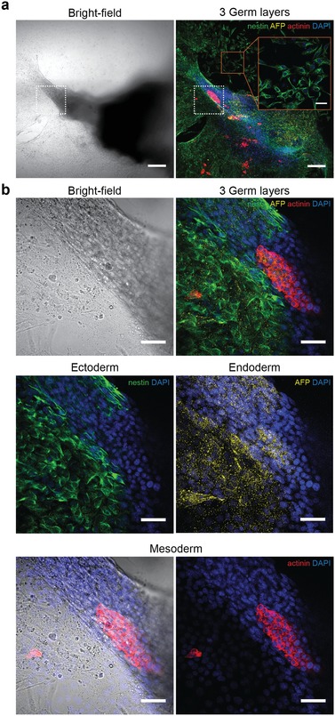
Immunofluorescent characterization of embryoid‐body differentiation. a) Bright‐field and confocal image of a spread‐out EB at 10× magnification, showing the spatial organization of the three germ layers at day 10. Ectodermal cells were stained with the neural‐progenitor marker nestin (green), endodermal cells with the carrier molecule alpha‐feto protein (AFP, yellow), and mesodermal cells with sarcomeric alpha actinin (red). The nuclear stain DAPI (blue) demonstrates the localization of the cells. A magnified area on the substrate surface (red frame) shows a monolayer of spread neural progenitor cells (scale bar: 50 µm). The area of contraction is indicated with a white frame. Scale bar: 200 µm. b) Bright‐field and confocal images of the area of contraction, indicated with the white frame in (a), at 40× magnification. Ectodermal cells, found at the periphery of the tissue, spread over the surface and formed monolayers of early neural progenitor cells. The endodermal marker AFP is scattered over the EB outgrowth zone, showing highest accumulation in AFP‐producing cells. Mesodermal cells were found in the center region of the EB, overlapping with the region of contraction (Figure 5b) as demonstrated in the bright‐field overlay. Scale bar: 50 µm.
