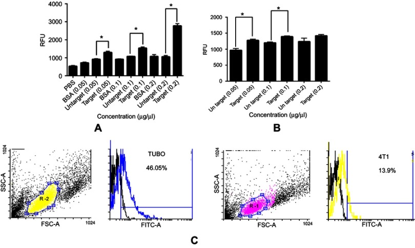
Figure 3.
Fluorimetry and flow cytometry analysis of binding ability of targeted and untargeted exosomes. Binding of PKH-67 labeled targeted and untargeted exosomes to (A) coated HER2 protein and (B) HER2-positive SKBR3 cells with different concentrations. Fluorescence of each well was assessed by fluorimetry after removing unbound exosomes. BSA and PBS were used as controls. (B) Binding of PKH-67 labeled targeted and untargeted exosomes to HER2-positive SKBR3 cells. There were significant differences both in HER2 protein and HER2-positive cell immobilization; yet, no significant difference was observed at 0.2 µg/µL in binding to SKBR3 cells. (C) Binding and entrance of targeted labeled exosomes were quantified by flow cytometry. The results show a significant increase in binding of targeted exosomes to TUBO cells (46.05%) as HER2-positive murine cells compared to 4T1 cells (13.9%). Targeted exosomes showed significantly higher binding to HER2-positive cells compared to HER2-negative cells. Each error bar represents the mean±SD of three replicates. *P<0.05.
Abbreviations: RFU, relative fluorescence units; BSA, bovine serum albumin.

