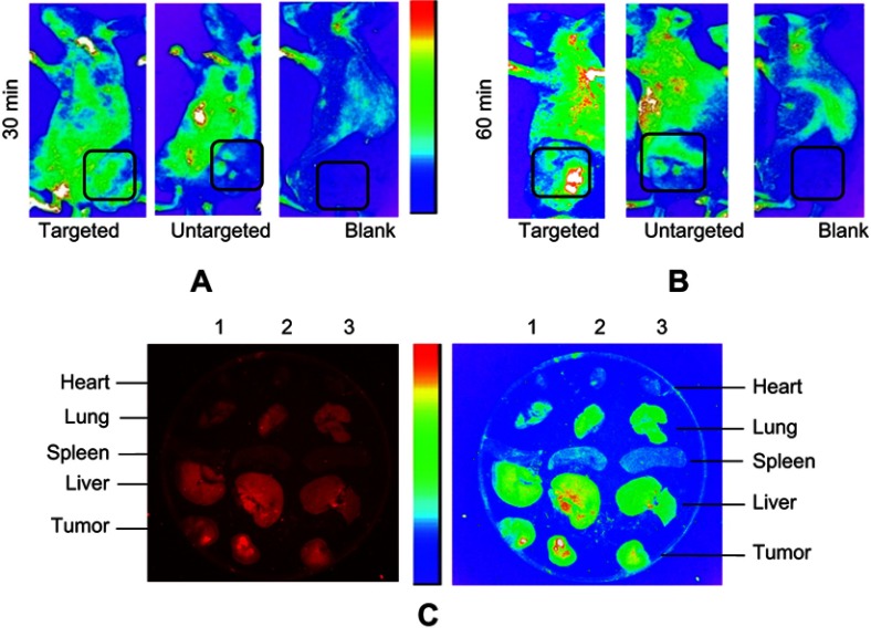
Figure 7.
Biodistribution of exo-DOX in mice bearing TUBO tumor. In vivo targeting efficiency of targeted exo-DOX were assessed in a murine TUBO tumor model. These models were administered with a single 70-µg injection of targeted or untargeted exo-DOX. (A) In vivo fluorescent signals were recorded at 30 minutes and (B) 60 minutes post-injection. Fluorescence was detected at the tumor sites (black boxes), with gradual increasing in signal intensity at 60 minutes. Enhanced permeability and retention (EPR) effect causes accumulation of untargeted exo-DOX in tumor site, while it was lower in comparison to targeted exo-DOX. PBS was injected as blank and no fluorescence was detected in these mice. (C) Ex vivo fluorescent imaging of major organs of the tumor model after 60 minutes. Lanes 1, 2, and 3 were sequentially related to untargeted, targeted, and PBS intravenous injections. Accumulation of targeted and untargeted exo-DOX was mainly in the liver and lung, while, there was no accumulation in the heart after 60 minutes. Fluorescent intensity of untargeted exo-DOX was lower than targeted exo-DOX.
Abbreviations: DOX, doxorubicin; exo-DOX, doxorubicin-loaded exosome DOX.

