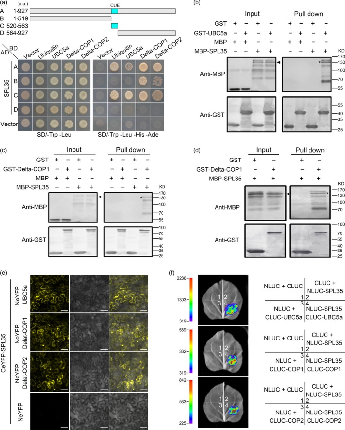
Figure 6.

Interaction of SPL35 with OsUBC5a, Delta‐COPI and Delta‐COP2. (a) Yeast two‐hybrid assays for interaction of SPL35 with Ubiquitin, OsUBC5a, Delta‐COP1 or Delta‐COP2. The full‐length and different fragments of SPL35 were inserted to the prey (AD) vector pGADT7, and the ubiquitin, OsUBC5a, Delta‐COP1 and Delta‐COP2 were inserted into vector pGBKT7 as baits. Yeast strains were cultured on the SD/–Trp‐Leu and SD/–Trp‐Leu‐His‐Ade selection medium with 3 mm 3‐amino‐1,2,4‐triazole. (b–d) Pull‐down assays for the interactions of SPL35 with OsUBC5a, Delta‐COP1 and Delta‐COP2 in vitro respectively. GST (26.30 kD), GST‐OsUBC5a (42.87 kD), GST‐ Delta‐COP 1 (84.07 kD), GST‐ Delta‐COP2 (84.07 kD), MBP (42.5 kD) and MBP‐SPL35 (143.25 kD) were expressed in bacteria. Pull‐down was performed using GST binding resin. Proteins were detected with antibodies as indicated. Similar results were obtained in three independent experiments. The asterisk shows the band pulled down, and the arrow shows the immune‐blotting band of MBP‐SPL35 by the anti‐MBP monoclonal antibody. (e) Interactions between SPL35 and OsUBC5a, Delta‐COP1 and Delta‐COP2 shown by BiFC assays in Nicotiana benthamiana leaf epidermal cells. BiFC fluorescence is indicated by the eYFP signal. The signal of eYFP was not detected in the corresponding negative controls (fourth panels). eYFP, enhanced yellow fluorescent protein fluorescence; DIC, differential interference contrast. Scale bars, 10 μm. (f) firefly LUC complementation imaging (LCI) assay detecting the interaction between SPL35 and OsUBC5a, Delta‐COP1 and Delta‐COP2. The coloured scale bar indicates the luminescence intensity. NLUC indicates the N terminus of LUC, while CLUC indicates the C terminus of LUC.
