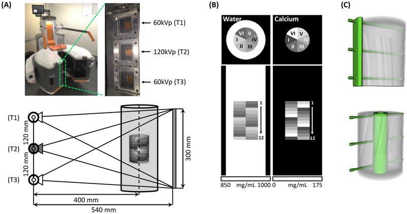
Fig.1.
(A) Extremity CBCT system with an axial multi-source x-ray unit. System geometry and source firing sequence for a single-scan DE acquisition is shown. (B) Digital multi-material phantom (axial and coronal view) used in validation studies. The phantom consisted of an outer water container with an inner insert containing water-Ca mixtures of varying Ca concentration. (C) The placement of metal implants (top: plate, bottom: intramedullary nail) with respect to the inner insert of the phantom.

