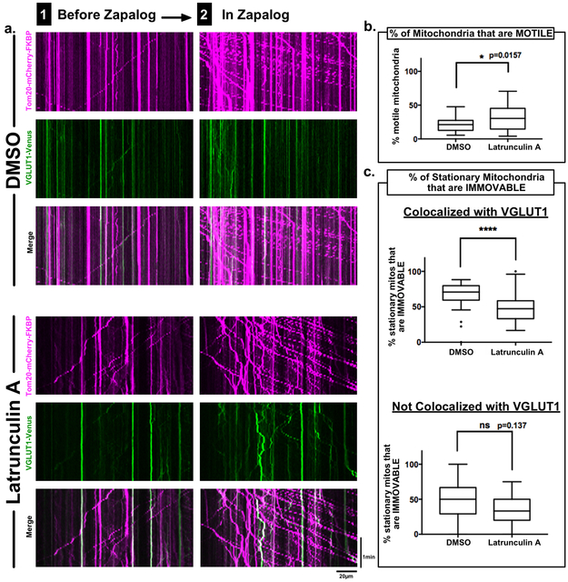
Figure 5. Actin contributes to anchoring of mitochondria at synapses.
a, Axonal kymographs from live-imaging of neurons co-transfected with Kif1a(1-489)-DHFR-Myc, Tom20-mCherry-FKBP and the presynaptic marker VGLUT1-Venus, incubated for 6hrs with either DMSO or 2.5μM Latrunculin A, and imaged before (1) and after (2) addition of 1μM zapalog.
b, Quantification of the percentage of mitochondria that are motile before addition of zapalog in neurons incubated in DMSO vs neurons incubated with Latrunculin A.
c, Quantification of the percentage of mitochondria that are anchored, i.e. remain stationary even after addition of zapalog, subdivided into two groups: those that colocalize with VGLUT1-Venus and those that do not colocalize with VGLUT1-Venus.
n = 28 axons ea / 4 independent repeats; box-and-whisker plots display statistical median (center value), upper and lower quartiles (boxes), max values (whiskers), and outliers (dots); KS nonparametric tests; ns = p>0.05, * = p≤0.05, **** =p≤0.0001, source data in Sup. Table 1.

