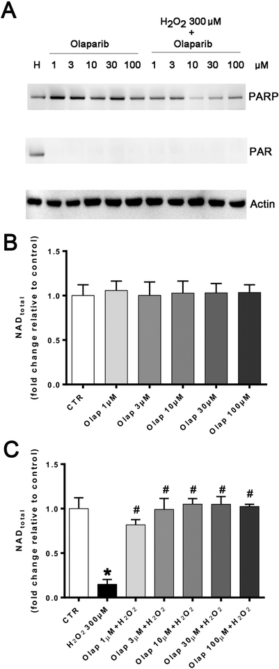
Figure 24. Effect of olaparib on PARP activation and cellular NAD+ levels in U937 cells subjected to H2O2-induced oxidative stress in vitro.
(A): Western blotting for PARP1 (top lanes), polyADP-ribose (PAR), the enzymatic product of PARP activation (middle lanes) and actin loading control (bottom lanes) in U937 cells treated with olaparib (1, 3, 10, 30 or 100 μM) in control and in 300 μM H2O2 (“H”) challenged cells at 1 hour. Note the increase in PAR (indicative of PARP activation) in response to oxidative stress, and the full inhibition of this response at all olaparib concentrations tested. The expression of PARP1 was unaffected by olaparib in any of the control conditions tested. However, in the oxidatively stressed cells, in the presence of olaparib, the expression of PARP1 also appeared to be downregulated. Western blots shown are representative of experiments performed on 3 different experimental days. (B): Effect of olaparib on the cellular NAD+ levels in control cells (without oxidative stress challenge). Cells were treated with olaparib (1, 3, 10, 30 or 100 μM) for 1 hour. Data are shown as mean±SEM of n=3 determinations. (C): Effect of olaparib on the cellular NAD+ levels in cells subjected to oxidative (H2O2). Cells were treated with olaparib (1, 3, 10, 30 or 100 μM), followed immediately by 300 μM H2O2 for 1 hour. Data are shown as mean±SEM of n=3 determinations. *p<0.05 shows significant decrease in NAD+ levels in response to H2O2, compared to control cells; #p<0.05 shows significant protective effect of olaparib in H2O2 challenged cells compared to vehicle-treated H2O2 challenged cells.

