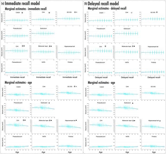
Figure 1.
Marginal estimates of hippocampal subfield volumes (mm3) from immediate recall (IR) and delayed recall (DR) models, showing effects of IR, DR and age. (a) Upper panels present marginal estimates (blue line; light blue band ±95% CI) of subfield volumes as a function of IR score; overlaid cyan scatter presents observed participant-wise data. Lower panels present marginal estimates (±95% CI) of subfield volumes as a function of age (years), with participant-wise scatters. (b) Marginal estimates for DR model; all specifications as per (a). *IR/DR cubic term significant, p < 0.05; • age quadratic term significant, p < 0.05; grey shading denotes marginally significant trend - see Tables 2 and 3, and Results. Note differences in y-axis ranges across panel rows in (a,b); adjusted to accommodate differences in subfield volumes (see also Supplementary Fig. 2). All marginal estimates calculated from fully-adjusted models, holding all covariates at their means.

