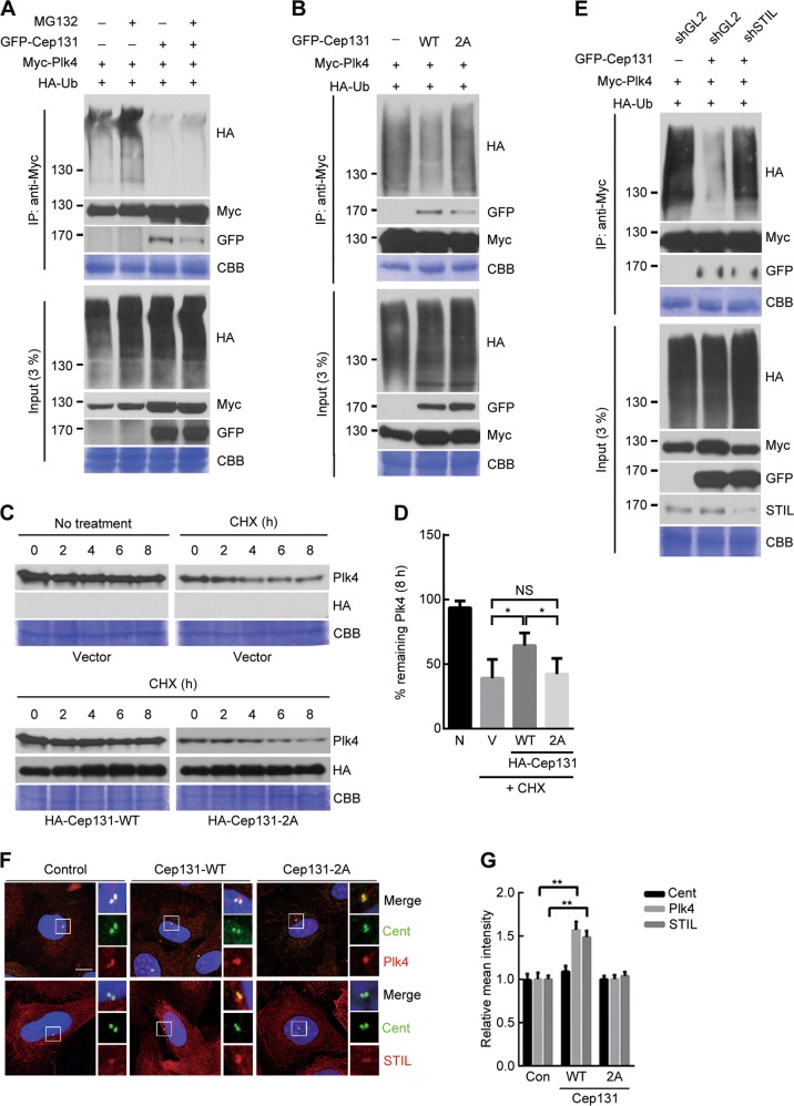
Fig. 6. Cep131 overexpression induces Plk4 stabilization and STIL accumulation at the centriole.
a, b HEK293T cells co-expressing Myc-Plk4 and HA-Ub and/or GFP-Cep131 were treated with MG132 for 6 h and immunoprecipitated. Ubiquitylation properties were analyzed by immunoblotting. c U2OS cells with doxycycline-inducible expression of Myc-Plk4 were treated with 10 μM cycloheximide (CHX) for 8 h. Every 2 h, cells were harvested and analyzed by immunoblotting. d Quantification of Plk4 band intensity measured with ImageJ. Error bars represent means ± SD from three independent experiments. **P < 0.01, unpaired Student’s t test. NS, not significant. e HEK293T cells stably expressing shGL2 (control) or shSTIL were transfected with HA-Cep131-WT or 2A and immunoprecipitated. Ubiquitylation properties were analyzed by immunoblotting. f Immunofluorescence analysis of excessive accumulations of Plk4 (top panel) and STIL (bottom panel) at the centriole in U2OS cells transfected with Cep131-WT or 2A. Insets are approximately fivefold magnified at the centrosomal region. Scale bar, 10 μm. g Quantification of fluorescence intensity of Cent (left), Plk4 (middle), and STIL (right) at the centriole. Over 50 centrioles were measured for each condition. **P < 0.01, unpaired Student’s t test. CBB staining use as a loading control in a, b, c, e

