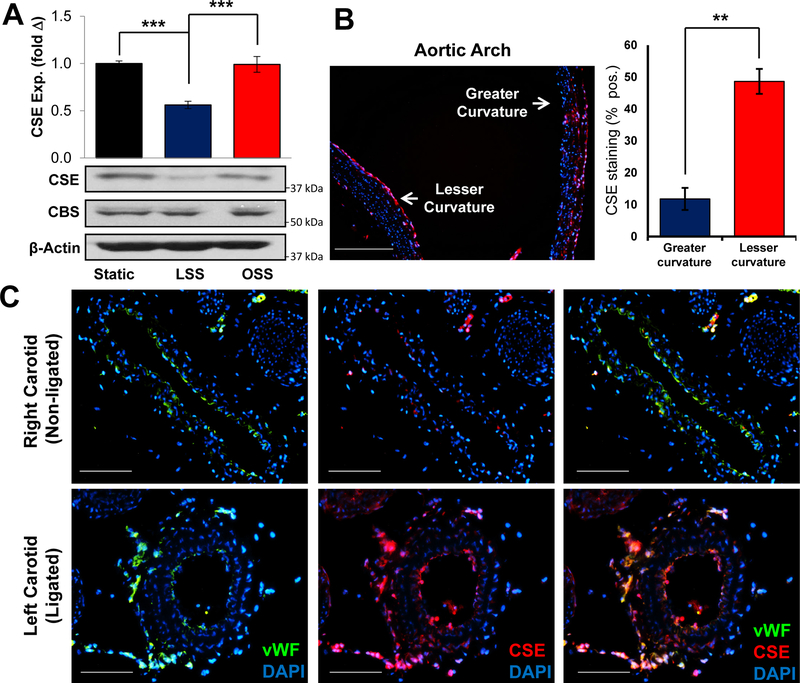
Figure 1. Shear stress regulates CSE expression.
Human aortic endothelial cells (HAECs) were exposed to LSS or OSS for 16 hours. (A) CBS and CSE expression was assessed by Western blotting. Representative images are shown. (n=5, *** p<0.001 by One-Way ANOVA). CSE protein expression was assessed by immunostaining at sites of laminar (greater curvature) or disturbed flow (lesser curvature). (scale bar = 200 μm, n=5, ** p<0.01 by Mann-Whitney U test). (C) Partial carotid ligation was performed in WT mice to induce disturbed flow in the ligated left carotid compared to the unligated right carotid. (scale bar = 100 μm, n=7).

